



: To examine the effects of 12 weeks of strength and resistance training on serum calcium, phosphorus, and parathyroid hormone (PTH) in elite under-23 (U23) male footballers, and to elucidate differential mechanobiological and endocrine mechanisms underlying distinct training modality responses, a question previously unaddressed in this population. Methods: Thirty elite U23 male footballers were assigned to a resistance training group (RTG; n = 10), strength training group (STG; n = 10), and control group (CG; n = 10) in a quasi-experimental, pretest–posttest design. RTG and STG completed supervised training three times per week for 12 weeks while CG continued routine football training. Fasting venous blood samples were collected pre and post intervention to assess serum calcium, phosphorus, and PTH, and analysed with one-way ANOVA and Tukey's HSD. Results: Post intervention ANOVA revealed highly significant between-group differences for calcium (F(2,27) = 131.01, η²p = .91), phosphorus (F(2,27) = 74.91, η²p = .85), and PTH (F(2,27) = 54.35, η²p = .80) (all p < .001). RTG elicited significantly higher serum calcium than STG (mean difference = 1.03 mg/dL, p < .001), while phosphorus and PTH responses were statistically comparable between training groups. Conclusion: Twelve weeks of structured training significantly improved mineral homeostasis and suppressed PTH in elite U23 footballers. Resistance training elicited a superior calcaemic response, likely attributable to greater cumulative mechanical strain volume. Routine football training alone failed to induce comparable biochemical adaptations, highlighting the need for structured supplementary loading in elite football conditioning.
Football (soccer) is among the most mechanically demanding team sports, exposing the skeleton to high magnitude, multidirectional forces through repeated sprinting, jumping, tackling, and rapid directional changes. These loading patterns generate potent osteogenic stimuli via mechanotransduction, promoting bone modelling and adaptation that supports superior bone mineral density (BMD) and structural properties relative to non-weight-bearing activities.1 This skeletal benefit is especially significant during late adolescence and early adulthood, the primary window for peak bone mass accrual, when exercise induced strain augments bone mineral content (BMC) and cortical geometry at weight bearing sites such as the femoral neck and lumbar spine.1,2 Maximising peak bone mass during this developmental window is widely regarded as one of the most effective long term strategies for preventing osteoporosis and fracture in later life.3
In elite under 23 (U23) male footballers, musculoskeletal development during this life stage remains highly responsive to mechanical stimuli, creating a critical opportunity to simultaneously enhance skeletal integrity and athletic performance. However, as athletes accumulate years of sport specific training, their skeletons may reach a threshold of adaptation to habitual football loading, potentially reducing the osteogenic efficacy of routine training alone a phenomenon known as the 'training saturation' effect.4 In this context, the supplementary mechanical stimulus provided by structured resistance or strength training may be necessary to continue driving beneficial adaptations in bone mineral homeostasis and endocrine regulation. Despite the growing recognition of skeletal health in elite sport, whether routine football training is sufficient to optimise biochemical markers of bone metabolism in U23 athletes, and how specific supplementary training modalities differentially influence these markers, remain poorly characterised questions in the literature5,6
The endocrine regulation of bone metabolism particularly the interplay between serum calcium, phosphorus, and parathyroid hormone (PTH) represents a critical and often overlooked dimension of skeletal adaptation to exercise. PTH acts as the primary guardian of systemic calcium homeostasis, regulating bone resorption, renal calcium reabsorption, and intestinal calcium absorption through vitamin D activation.7,8 Acute and chronic exercise induced perturbations in serum calcium and phosphorus directly modulate PTH secretion, creating a dynamic interaction between mechanical loading and endocrine bone regulation.7 Characterising how different training modalities influence this mineral endocrine axis is therefore essential for understanding the full scope of exercise induced skeletal adaptation in elite athletes.
While previous research has broadly documented osteogenic effects of resistance training across general populations, direct comparative evidence contrasting moderate intensity, higher volume resistance training against high load, low volume strength training with respect to serum calcium, phosphorus, and PTH is absent in elite U23 footballers. This represents a specific and important scientific gap: if training volume rather than peak intensity is the primary determinant of calcaemic adaptation as mechanobiological theory predicts, then resistance training should elicit a superior biochemical response compared with strength training; yet this hypothesis has not been empirically tested in an elite team sport context.9,10 The present study was therefore designed to fill this gap through a controlled quasi experimental trial. It was hypothesised that both training modalities would significantly improve mineral homeostasis and suppress PTH relative to routine football training, with resistance training producing a superior calcaemic response attributable to its greater cumulative mechanical loading volume.
Literature reviewTheoretical framework: mechanotransduction and bone adaptationThe biomechanical basis of bone adaptation is grounded in two complementary theoretical frameworks. Julius Wolff's law (1892) established the foundational principle that bone architecture adapts structurally in response to the habitual mechanical loads imposed upon it, with bone deposited along lines of mechanical stress and resorbed where loading is minimal. Harold Frost subsequently formalised this concept into the mechanostat theory, proposing that bone is governed by a feedback control system in which specific strain magnitude thresholds the minimum effective strain (MES) trigger either bone modelling (net gain) or remodelling (maintenance or loss).9,11 Frost's model further stipulated that not only strain magnitude, but also rate, frequency, and cumulative cycle number determine the osteogenic response, with higher frequency, lower magnitude loading potentially producing greater cumulative osteogenic signalling than fewer, higher magnitude strain events a principle with direct implications for comparing resistance versus strength training.10
Contemporary mechanobiology has substantially expanded Frost's original framework, identifying the molecular pathways through which mechanical strain is transduced into cellular bone forming signals. Osteocytes, embedded within the bone matrix, act as primary mechanosensors, detecting fluid shear stress generated by strain induced canalicular flow and transmitting anabolic signals via prostaglandin E2, nitric oxide, and Wnt/β catenin pathway activation.12 Mechanical loading also suppresses sclerostin a Wnt inhibitor produced by osteocytes thereby disinhibiting Wnt driven osteoblast differentiation and bone matrix synthesis. Simultaneously, loading modulates the RANKL/OPG ratio, reducing osteoclastogenesis and bone resorption.13,14 These cellular mechanisms collectively link mechanical strain to net bone formation, providing the molecular basis for exercise induced increases in BMD and mineral content documented across intervention studies.
A critical limitation of applying mechanostat theory to exercise intervention research is that most supporting mechanistic evidence derives from animal models or in vitro experiments, while human exercise studies typically measure structural outcomes (BMD via DXA) or indirect biochemical markers rather than directly quantifying strain or osteocyte signalling.15,16 The translation of mechanostat thresholds to specific training parameters (sets, repetitions, intensity) in human athletes therefore remains largely inferential, and the relative osteogenic potency of different resistance training configurations—particularly moderate volume versus high load protocols has not been definitively established through biochemical biomarker studies in elite sport contexts. The present study addresses this gap by using serum calcium, phosphorus, and PTH as surrogate biochemical markers of the mineral homeostasis response to differential training volume loading in elite U23 footballers.
Football, mechanical loading, and bone healthFootball participation has been consistently associated with superior bone characteristics compared with non-weight bearing sports and sedentary controls, with recreational and elite footballers demonstrating significantly higher BMD, BMC, and cortical bone geometry at weight bearing sites including the tibia, femoral neck, and lumbar spine.17,18 A systematic review and meta-analysis by1 confirmed that even recreational football participation for 12–65 weeks produced meaningful BMD improvements, particularly at the femoral neck and total hip, in both men and women across a wide age range. These structural adaptations are attributed to the multidirectional, high impact loading patterns inherent in football sprinting, jumping, and rapid deceleration that generate peak ground reaction forces sufficient to exceed osteogenic strain thresholds at weight bearing skeletal sites.1
However, despite well documented structural benefits, evidence regarding the effects of football participation on biochemical markers of bone metabolism particularly the calcium phosphorus PTH mineral axis remains sparse and inconsistent.5 observed that even elite academy soccer players from the English Premier League exhibited markers of bone resorption elevation under high volume training conditions with reduced carbohydrate availability, suggesting that habitual football training does not automatically ensure favourable mineral homeostasis.2 reported that sport type and biological sex modulated bone turnover markers in young athletes, with football players demonstrating distinct biochemical profiles compared with swimmers and track athletes, yet specific calcium phosphorus PTH data in elite U23 male footballers remained absent from that analysis. Critically, whether the osteogenic loading inherent in routine elite football training is sufficient to maintain optimal biochemical markers of mineral homeostasis in U23 athletes who have already achieved a degree of skeletal adaptation through years of competition has not been directly investigated constituting a central justification for the present study.
The unique musculoskeletal demands of elite level U23 football also introduce specific skeletal health considerations not fully addressed in recreational football research. Elite U23 players undergo substantially higher training volumes, intensities, and match densities than recreational counterparts, which may simultaneously create greater osteogenic stimuli and elevated risk of bone stress injuries, fatigue fractures, and training induced mineral depletion;19Marriott et al., 2024). The adolescence to adulthood transition period characteristic of U23 football also coincides with the final phases of peak bone mass consolidation, during which training induced adaptations can have long lasting consequences for skeletal integrity and injury resilience throughout an athletic career.3 These factors collectively highlight the importance of characterising the biochemical dimensions of bone metabolic health in this specific population, beyond the structural BMD outcomes that dominate existing football bone literature.
Resistance and strength training: effects on bone metabolismProgressive resistance training is among the most evidence supported exercise modalities for improving bone metabolic health across diverse populations. Systematic reviews and meta analyses consistently demonstrate that resistance training programs incorporating moderate to high intensities (≥60–70% 1RM), multi joint exercises, and progressive overload are associated with significant improvements in BMD at the lumbar spine, femoral neck, and total hip in adults, older adults, and postmenopausal women20–22 demonstrated that even short term (12 week) heavy resistance training produced significant improvements in bone formation markers in well-functioning older adults, while23 confirmed in a comprehensive meta-analysis that resistance exercise was among the most effective interventions for improving both BMD and bone turnover markers in adults across a broad age spectrum.
The mechanisms through which resistance training promotes bone adaptation extend beyond simple mechanical loading to include hormonal and endocrine pathways. Resistance training acutely elevates anabolic hormones including growth hormone (GH), insulin like growth factor 1 (IGF 1), and testosterone which exert direct stimulatory effects on osteoblast proliferation and bone matrix synthesis.24,25 Chronically, resistance training has been shown to attenuate basal PTH levels through enhanced calcium availability, reducing the endocrine drive for bone resorption and shifting the bone metabolic environment towards net formation7,26,27 demonstrated in a sixeen-month resistance training intervention that PTH was significantly reduced alongside improvements in calcium metabolism, without requiring changes in vitamin D status suggesting that mechanical loading per se can drive favourable PTH modulation independent of nutritional supplementation.28 Similarly observed significant reductions in PTH and increases in serum calcium and phosphorus following a structured exercise programme in older adults, supporting the view that exercise induced endocrine adaptations contribute to improved mineral homeostasis across populations.
High load strength training characterised by heavier intensities (≥75–90% 1RM) and lower repetition volumes has well established neuromuscular benefits including superior maximal strength gains and neural adaptations, but its specific effects on biochemical markers of bone mineral metabolism are less thoroughly investigated than moderate volume resistance training.29,30 The mechanostat framework predicts that high load strength training, by generating larger peak strain magnitudes at lower cycle frequencies, may produce a different osteogenic signal profile than moderate load, higher volume resistance training potentially eliciting a less pronounced cumulative calcaemic adaptation despite comparable or superior neuromuscular stimulation.24 emphasised that the importance of muscular strength for athletic performance does not necessarily align with optimal osteogenic stimulation, as the loading configurations most effective for strength development (high intensity, low volume) may not maximise cumulative strain cycles at bone tissue surfaces. This mechanobiological distinction between training volume and intensity effects on the calcium phosphorus PTH axis represents a critical and underexplored scientific question addressed by the present study.
Calcium, phosphorus, and PTH regulation: exercise interactionsCalcium and phosphorus are the primary inorganic constituents of bone, with approximately 99% of body calcium and 85% of body phosphorus stored in the skeleton as carbonated hydroxyapatite [Ca10(PO4)6(OH)2] the crystalline mineral phase that confers compressive strength and structural rigidity to the bone matrix.8,31 Systemic calcium homeostasis is maintained within a narrow physiological range (8.5–10.2 mg/dL in adults) through a tightly regulated hormonal triad: PTH stimulates bone resorption and renal calcium reabsorption while activating the renal conversion of 25 hydroxyvitamin D to calcitriol (1,25 dihydroxyvitamin D3), which enhances intestinal calcium and phosphorus absorption; calcitonin inhibits osteoclast activity to suppress calcium efflux from bone; and FGF 23, produced by osteocytes in response to phosphate excess and mechanical loading, promotes renal phosphate excretion and suppresses calcitriol synthesis.8,32,33
Exercise induced perturbations in systemic mineral balance create a complex and dynamic interaction between mechanical loading and endocrine bone regulation. Acute exercise transiently reduces ionised serum calcium through increased skeletal calcium uptake during bone remodelling and muscle contraction related calcium flux, triggering a compensatory rise in PTH that restores calcium homeostasis by stimulating bone resorption and renal reabsorption.7 The magnitude and duration of this PTH response depend critically on exercise type, intensity, and duration: high intensity anaerobic exercise tends to produce transient PTH spikes that may have anabolic bone effects, whereas prolonged endurance exercise is associated with sustained PTH elevation favouring net bone resorption a distinction relevant to comparing the football conditioning context with supplementary resistance or strength training.7,34
Chronic adaptation to structured resistance training appears to shift the setpoint of mineral homeostasis regulation, attenuating basal PTH and elevating fasting serum calcium and phosphorus through mechanisms involving enhanced intestinal absorption efficiency potentially mediated by upregulated calcitriol synthesis improved renal calcium retention, and reduced bone resorption activity.7,33,35observed that young male athletes competing in different sports demonstrated significantly different calcium, phosphate, and PTH profiles, with sport specific mechanical loading patterns accounting for a substantial proportion of endocrine variation.36 demonstrated that exercise induced vitamin D and calcium metabolism adaptations were apparent within 12 weeks in military recruits undergoing structured physical training, suggesting that the duration of the present intervention (12 weeks) is sufficient to detect meaningful biochemical adaptations in a well-controlled cohort. Critically, however, the majority of existing evidence on exercise mineral homeostasis interactions derives from non athlete populations or uses acute post exercise measurement designs rather than controlled chronic intervention comparisons creating the specific gap in evidence that motivates the present study.
Evidence gaps and rationale for the present studySynthesising the literature reviewed above reveals several interconnected gaps that collectively justify the design and objectives of the present investigation. First, while the osteogenic structural effects of football participation are well documented, the specific effects of routine elite football training on biochemical markers of mineral homeostasis serum calcium, phosphorus, and PTH in U23 male athletes have not been characterised in controlled studies. Second, although resistance training is broadly associated with improved bone metabolic markers, direct comparisons between moderate volume resistance training and high load strength training on the calcium phosphorus PTH triad in elite team sport athletes are absent from the literature. Third, the mechanobiological hypothesis that training volume rather than peak load intensity is the primary driver of calcaemic adaptation in humans has theoretical support but lacks direct empirical testing in elite athlete populations using biochemical biomarkers. Fourth, the U23 developmental stage during which peak bone mass consolidation is ongoing and supplementary training may have lasting skeletal consequences has received insufficient attention in exercise bone adaptation research relative to older adult, paediatric, or non-athlete populations.37
The present study addresses all four gaps through a controlled quasi experimental comparison of resistance training, strength training, and routine football training in elite U23 male footballers, with serum calcium, phosphorus, and PTH as biochemical outcome markers. This integrated design enables, for the first time, direct modality comparison of training effects on the mineral endocrine axis in this population, with findings interpretable against both mechanobiological theoretical frameworks and existing exercise bone intervention evidence. The results contribute novel, population specific evidence to the intersection of sport science, bone physiology, and endocrinology, with direct implications for evidence-based exercise prescription in elite football development programmes.
MethodsStudy designThis study was conducted as a quasi-experimental, non randomised, pretest–posttest controlled group trial, adhering to the updated CONSORT 2025 guidelines and the TREND statement for non randomised behavioural interventions, to evaluate the effects of a 12 week resistance and strength training intervention on biochemical markers of bone metabolism in elite U23 male football players.38 The trial was registered with the Directorate General of Sports, Khyber Pakhtunkhwa under the first registration date on 27/06/2019.The quasi experimental design was selected given the logistical and ethical constraints of assigning elite athletes from club environments to fully randomised conditions a limitation acknowledged as a potential source of selection bias, mitigated through stratified group allocation and baseline equivalence verification.39,40 While quasi experimental designs preclude definitive causal inference at the level of a fully randomised controlled trial, the use of stratified allocation, a concurrent control group, and verified baseline equivalence strengthens internal validity within the practical constraints of elite sport research.
ParticipantsThirty elite football players affiliated with clubs in the Peshawar Division of Pakistan volunteered to participate. All had a minimum of three years of structured football training and met predefined eligibility criteria: absence of musculoskeletal injuries, metabolic bone disease, chronic illness, or use of medications or supplements known to influence bone metabolism. Participants were allocated to three groups resistance training (RTG; n = 10), strength training (STG; n = 10), and control (CG; n = 10) with assignments stratified by age, playing level, and baseline anthropometric characteristics (Hidayat et al., 2025). A priori power analysis using G*Power 3.1 confirmed that n = 10 per group provided >80% power to detect medium to large between group effects (Cohen's f = 0.40) at α = .05 for one way ANOVA, consistent with effect sizes reported in comparable exercise bone metabolism interventions.41 The sample size of n = 10 per group is acknowledged as a limitation; future studies should employ larger samples to improve statistical power and generalisability.
Training interventionThe 12-week intervention comprised sessions conducted three times per week on non consecutive days. All sessions were supervised by certified strength and conditioning professionals to ensure technical correctness, safety, and dose fidelity. The RTG performed a moderate to high volume programme targeting major muscle groups (squats, lunges, leg press, chest press, lat pulldown, shoulder press, and core exercises) at 60–75% 1RM, with 3–4 sets of 10–12 repetitions per exercise and systematic progressive overload. The STG performed identical exercises at 75–90% 1RM with 3–5 sets of 4–6 repetitions per exercise and 2–3 min inter set rest to maximise force production, consistent with contemporary strength development principles.29,24,30 The CG continued regular football training without additional structured resistance or strength training. Session RPE (Borg CR 10 scale) was recorded post session and attendance registers were maintained. Mean adherence was 94.3% ± 3.8% for RTG and 93.7% ± 4.1% for STG.
Biochemical and anthropometric assessmentsFasting venous blood samples were collected at baseline (08:00–09:00 h) and within one week following the 12-week intervention to minimise diurnal variation. Serum concentrations of calcium (mg/dL), phosphorus (mg/dL), and PTH (pg/mL) were analysed using commercially available assay kits at a certified clinical laboratory following standardised manufacturers' protocols.42 Intra assay and inter assay coefficients of variation for all analytes were <5%. Anthropometric assessments included height, body mass (BM), BMI, waist circumference (WC), hip circumference (HC), and waist to hip ratio (WHR) following standard protocols.43,44 Three day dietary records were collected pre and post intervention and analysed using dietary analysis software to confirm comparable macronutrient and micronutrient (calcium, phosphorus, vitamin D) intake across groups.45
Statistical analysisData were analysed using SPSS version 26.0 (IBM Corp., Armonk, NY, USA). Descriptive statistics are presented as mean ± SD. Normality was assessed using the Shapiro Wilk test. Within group changes were evaluated using paired samples t tests and between group differences via one way ANOVA. When significant omnibus effects were detected, Tukey's HSD post hoc test identified pairwise comparisons. Statistical significance was set at p < .05.46,47,48 Effect sizes are reported as partial eta squared (η²p) for ANOVA (small = .01, medium = .06, large = .14) and Cohen's d for paired comparisons.
Ethical approvalThe study protocol was approved by the Institutional Research Ethics Committee and conducted in accordance with the Declaration of Helsinki.49 All participants provided written informed consent following verbal and written explanation of objectives, procedures, risks, and benefits. Participation was voluntary, and confidentiality was maintained throughout.50,51
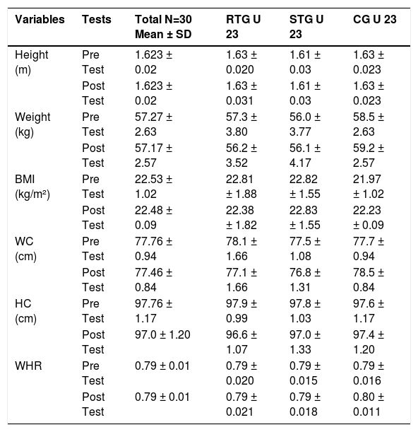
ResultsAnthropometric characteristicsBaseline anthropometric measurements for RTG, STG, and CG are presented in Table 1. No significant between group differences were detected at baseline for any variable (all p > .05), confirming initial group equivalence a critical prerequisite for valid between group comparisons in quasi experimental designs. Post intervention, no meaningful changes in anthropometric parameters were observed within any group, consistent with the short intervention duration and absence of targeted nutritional manipulation.
Anthropometric measurements of all three groups (Mean ± SD).
Note. RTG = Resistance Training Group; STG = Strength Training Group; CG = Control Group; WC = Waist Circumference; HC = Hip Circumference; WHR = Waist to Hip Ratio. No significant between group differences at baseline or within group changes post intervention (all p > .05).
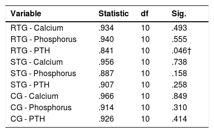
Shapiro Wilk normality test results for baseline serum calcium, phosphorus, and PTH are presented in Table 2. Serum calcium and phosphorus in all three groups, and PTH in STG and CG, were normally distributed (p > .05). PTH in the RTG showed a marginal deviation (p = .046); however, parametric tests were retained on grounds of established robustness to minor normality violations in samples of this size, consistent with methodological guidance.46,47
Shapiro wilk normality test results baseline biochemical variables.
Note. RTG = Resistance Training Group; STG = Strength Training Group; CG = Control Group. † Marginal deviation; parametric tests retained.46 All other variables p > .05.
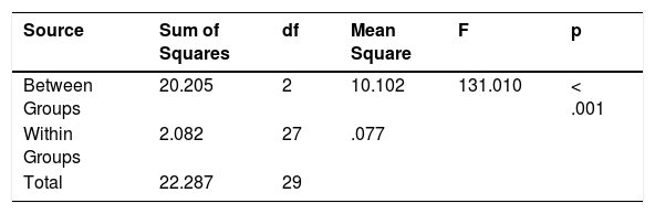
A one way ANOVA revealed a highly significant between group effect on post intervention serum calcium (F2,27 = 131.01, p < .001, η²p = .91) (Table 3), indicating that 91% of total post test calcium variance was attributable to group membership a very large effect by conventional benchmarks. Tukey's HSD post hoc analysis revealed a clear graded response: RTG > STG > CG. The mean difference between RTG and CG was 2.01 mg/dL (p < .001, 95% CI [1.70, 2.32]); between STG and CG was 0.98 mg/dL (p < .001, 95% CI [0.67, 1.29]); and between RTG and STG was 1.03 mg/dL (p < .001, 95% CI [0.72, 1.34]). All confidence intervals excluded zero, confirming robust pairwise differences. These calcium elevations, while remaining within the physiological reference range, represent clinically meaningful shifts in systemic mineral availability with potential significance for bone matrix mineralisation rate and osteoblast activity.8,33
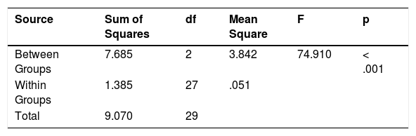
ANOVA results: serum phosphorusA significant between group effect was observed for post intervention serum phosphorus (F2,27 = 74.91, p < .001, η²p = .85) (Table 4). Post hoc comparisons confirmed that both RTG (mean difference = 1.17 mg/dL, p < .001, 95% CI [0.92, 1.42]) and STG (mean difference = 0.94 mg/dL, p < .001, 95% CI [0.69, 1.19]) differed significantly from CG. Crucially, no significant difference was observed between RTG and STG (mean difference = 0.23 mg/dL, p = .077, 95% CI [−0.02, 0.48]), indicating that both training modalities produced comparable beneficial effects on phosphorus homeostasis, in contrast to the calcium outcome. The p value of .077 for the RTG vs. STG phosphorus comparison approaches significance and warrants careful interpretation: with a larger sample, this comparison may have achieved statistical significance a limitation of the current sample size that future studies should address.
One eay ANOVA-Serum phosphorus post test.
| Source | Sum of Squares | df | Mean Square | F | p |
|---|---|---|---|---|---|
| Between Groups | 7.685 | 2 | 3.842 | 74.910 | < .001 |
| Within Groups | 1.385 | 27 | .051 | ||
| Total | 9.070 | 29 |
Note. η²p = .85 (very large effect). Post hoc Tukey HSD: RTG vs. CG (p < .001); STG vs. CG (p < .001); RTG vs. STG (p = .077, ns).
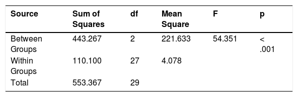
A significant main effect of group on post intervention PTH was found (F2,27 = 54.35, p < .001, η²p = .80) (Table 5). Post hoc analysis revealed that both RTG (mean PTH reduction = 8.80 pg/mL relative to CG, p < .001, 95% CI [6.56, 11.04]) and STG (mean reduction = 7.30 pg/mL, p < .001, 95% CI [5.06, 9.54]) demonstrated significantly lower PTH concentrations than CG. No significant difference in post test PTH was observed between RTG and STG (mean difference = 1.50 pg/mL, p = .238, 95% CI [−0.74, 3.74]), indicating that both training modalities produced comparable endocrine adaptations with respect to PTH suppression. The PTH reductions (7–9 pg/mL) are clinically meaningful given the narrow physiological range of PTH in healthy young adults and their potential downstream effects on bone resorption rate and turnover marker profiles.
One way ANOVA-parathyroid hormone post test.
| Source | Sum of Squares | df | Mean Square | F | p |
|---|---|---|---|---|---|
| Between Groups | 443.267 | 2 | 221.633 | 54.351 | < .001 |
| Within Groups | 110.100 | 27 | 4.078 | ||
| Total | 553.367 | 29 |
Note. η²p = .80 (very large effect). Post hoc Tukey HSD: RTG vs. CG (p < .001); STG vs. CG (p < .001); RTG vs. STG (p = .238, ns).
This study provides novel evidence that both resistance and strength training, when added to routine football conditioning, significantly and favourably modulate the calcium phosphorus PTH triad in elite U23 male footballers in contrast to routine football training alone, which failed to elicit comparable biochemical adaptations. These findings support the primary hypothesis and extend the exercise bone adaptation literature by providing the first population specific, controlled modality comparison evidence for mineral homeostasis responses in elite team sport athletes. The critical and clinically significant novel finding that resistance training produced a significantly superior calcaemic response compared with strength training, despite comparable PTH suppression supports the mechanobiological primacy of cumulative strain volume over peak strain intensity in driving calcaemic biochemical adaptations in this population. Each component of this finding warrants critical contextualisation against the theoretical frameworks, population specific factors, and methodological constraints described below.
The volume-dependent difference in serum calcium, with RTG showing significantly higher post-test calcium levels than STG, is consistent with mechanotransduction theory. This theory suggests that the cumulative number of strain cycles, rather than peak strain magnitude, plays a greater role in regulating osteoblast signaling and calcium mobilization.9 RTG's protocol of 10–12 repetitions at 60–75% 1RM generated substantially greater cumulative strain cycles per session than STG's 4–6 repetitions at 75–90% 1RM, providing a plausible mechanobiological basis for the superior calcium response. However, a critical methodological limitation is that cumulative volume loads (sets × reps × load) were not precisely quantified across individual participants throughout the 12 week period, preventing empirical dose response analysis linking specific loading parameters to biochemical outcomes. This limitation substantially constrains mechanistic interpretation and highlights the need for detailed training load documentation in future exercise bone intervention studies, ideally using velocity based tracking or session RPE load calculations (Hind & Burrows, 2022;.24 Additionally, serum calcium elevations may partly reflect enhanced intestinal calcium absorption mediated by exercise stimulated calcitriol synthesis a pathway that was not assessed in the present study due to the absence of 1,25 dihydroxyvitamin D measurements, constituting an important unmeasured mediator that future studies should characterise.33
The comparable phosphorus responses between RTG and STG despite divergent calcium outcomes likely reflect the tighter hormonal regulation of phosphate homeostasis via the PTH FGF 23 vitamin D axis, which maintains serum phosphorus within a narrow range independently of the specific training stimulus.8,32 In young healthy athletic cohorts, a potential ceiling effect in phosphate regulation may mask training specific differences particularly if both training modalities provided sufficient mechanical and metabolic stimuli to maximally stimulate phosphate regulating mechanisms regardless of volume differences. Critically, FGF 23 a principal osteocyte derived phosphatonin increasingly recognised as a key mediator of exercise phosphate interactions was not measured in this study.33 Without FGF 23 data, it is impossible to determine whether exercise induced FGF 23 suppression contributed to observed phosphorus elevations or whether comparable phosphorus responses in RTG and STG reflected similar FGF 23 modulation through volume independent mechanisms. This is a significant unmeasured variable that substantially limits mechanistic conclusions regarding phosphate regulation and that future studies must address through comprehensive hormonal profiling.
The comparable PTH reductions in both training groups relative to CG indicate a generalised beneficial endocrine adaptation to structured mechanical loading that is not specifically volume dependent a finding consistent with prior evidence that chronic exercise attenuates basal PTH through enhanced calcium availability reducing the parathyroid gland's secretory stimulus.7,26 The observed PTH reductions (7–9 pg/mL) accompanied by increases in serum calcium and phosphorus are consistent with a shift toward a more favourable bone metabolic environment: elevated circulating calcium reduces the stimulus for PTH secretion by suppressing the parathyroid gland's calcium sensing receptor (CaSR) mediated response, thereby attenuating the catabolic influence of sustained PTH elevation on bone tissue.26,52 This mechanistically parallels findings by7 and,53 who independently observed significant PTH attenuation alongside calcium and phosphorus improvements following structured exercise interventions in clinical and older adult populations, respectively. A critical interpretive constraint in the present study is the measurement of PTH at only a single post intervention timepoint, which does not capture the temporal dynamics of PTH adaptation across the 12-week period. Given the known biphasic nature of exercise PTH responses with acute post exercise spikes potentially followed by chronic baseline reductions mid intervention biochemical assessments at weeks 4 and 8 would substantially improve mechanistic understanding of the kinetics of PTH adaptation to training.7
The absence of significant biochemical changes in the CG despite sustained high volume football participation challenges the assumption that routine elite football training is sufficient to optimise bone metabolic health in U23 athletes. This finding aligns with the training saturation hypothesis: athletes with multiple years of competition level football experience may have already maximised their skeletal adaptation to habitual football specific loading patterns, requiring novel supplementary stimuli to drive further biochemical adaptations.5,54 However, a critical methodological caveat is that the CG's routine football training was not systematically monitored or quantified in this study training session frequency, volume, intensity, and load distribution across the 12-week period were not recorded, precluding rigorous comparison of total mechanical stimuli across all three groups. This is a substantial design limitation because without quantifying the CG's football training load, it is impossible to definitively determine whether the absence of biochemical adaptation is due to training saturation or a genuine insufficiency of football-specific mechanical stimulus compared to structured resistance training. Future studies should implement comprehensive training load monitoring across all groups using validated objective methods, such as GPS tracking, session RPE load, and heart rate monitoring, to allow accurate cross-condition comparisons.
Several broader methodological limitations merit explicit acknowledgement. The quasi experimental, non randomised design introduces potential selection bias, as unmeasured confounders genetic predisposition to bone adaptation, habitual physical activity outside the protocol, psychological motivation, and sleep quality may have differentially influenced outcomes across groups despite stratified allocation. The sample size of n = 10 per group, while adequate for the observed large effect sizes, substantially limits statistical power for detecting modest effects and increases risk of both Type II error and overestimation of true effect sizes due to winner's curse bias in small sample research (Hind & Burrows, 2022). The exclusively male, Pakistani elite football cohort limits generalisability to female athletes, players from different cultural or climatic contexts, and athletes at different performance levels. The absence of DXA based BMD assessments precludes determination of whether observed biochemical adaptations translated into structural skeletal changes arguably the most clinically important outcome from a long term bone health perspective.3 Finally, the biomarker panel was restricted to serum calcium, phosphorus, and PTH; the absence of specific bone formation markers (P1NP, osteocalcin) and resorption markers (CTX I) prevents characterisation of whether observed mineral changes reflected net bone formation or resorption, substantially limiting mechanistic interpretation (23)Sherrington et al., 2024). These limitations collectively indicate that the present findings should be interpreted as hypothesis generating and requiring replication in larger, more comprehensively designed trials before informing definitive clinical practice recommendations.
Practical applicationsThe findings carry direct evidence informed implications for strength and conditioning practitioners, sports scientists, sports physicians, and medical staff in elite U23 football programmes. The demonstration that routine football training fails to optimise bone related biochemical markers despite high sport specific training volumes underscores the necessity of incorporating structured supplementary resistance or strength training within elite football conditioning, particularly during the U23 developmental stage when peak bone mass consolidation remains biologically active.55,56 For practitioners prioritising skeletal health, the data suggest that moderate intensity, higher volume resistance training (60–75% 1RM, 10–12 repetitions) produces a superior calcaemic stimulus compared with high load strength training, consistent with the mechanostat principle of cumulative strain volume primacy. Undulating periodisation models that cycle between resistance emphasis (higher volume, moderate intensity) and strength emphasis (lower volume, higher intensity) phases may optimally stimulate both osteogenic and neuromuscular adaptive pathways without creating chronic overload.
Practitioners should note that while resistance training produced superior serum calcium elevation, both training modalities produced comparable and clinically meaningful PTH suppression indicating that high load strength training provides equivalent endocrine bone protection relative to resistance training. Coaches and sports scientists should therefore not exclusively privilege resistance training over strength training when designing conditioning programmes, particularly given that strength training's superior neuromuscular and maximal force production adaptations remain critical for football performance objectives.24,57 Implementing routine biochemical monitoring (serum calcium, phosphorus, PTH) during periods of intensified training can provide objective early indicators of bone metabolic stress, enabling proactive prescription adjustments before structural bone injury risk increases.58 All structured training interventions should be supported by evidence based nutritional strategies adequate dietary calcium (≥1000 mg/day), phosphorus, and optimised vitamin D status (serum 25 hydroxyvitamin D ≥50 nmol/L) to maximise the biochemical response to mechanical loading.33,45
ConclusionThis quasi experimental study provides novel, population specific evidence that 12 weeks of structured resistance or strength training significantly and beneficially modulates the calcium phosphorus PTH triad in elite U23 male footballers’ effects not replicated by routine football training alone. The key novel contribution is the empirical demonstration that resistance training (moderate intensity, higher volume) elicits a significantly superior calcaemic response compared with high load strength training, supporting the mechanobiological prediction that cumulative loading strain volume rather than peak load intensity is the primary driver of calcaemic biochemical adaptation in elite athletes. Comparable PTH suppression across both training modalities indicates that endocrine bone protection is achievable through either approach, with potential consequences for long term skeletal health and injury resilience in elite football.
Critically, the study's findings are bounded by important methodological constraints including a quasi-experimental design, small sample size, limited biomarker panel, absence of DXA based BMD assessment, unquantified CG training load, and single timepoint post intervention biochemical measurement. These limitations define the boundaries of causal inference that can be drawn from these data and identify clear priorities for future research:1 incorporate comprehensive bone metabolic panels (P1NP, CTX I, osteocalcin, FGF 23, 1,25 dihydroxyvitamin D);2 employ DXA based structural bone assessment;3 quantify training volume loads precisely across all groups;4 implement multi timepoint biochemical monitoring at weeks 0, 4, 8, and 12;5 enrol larger, more diverse, internationally representative samples; and6 use full randomisation to strengthen causal inference. The present findings constitute an empirically grounded foundation for this future research agenda, and support the practical recommendation that structured supplementary resistance or strength training be integrated into elite football conditioning programmes to promote mineral homeostasis and endocrine bone health in U23 athletes during the critical peak bone mass consolidation window.
FundingNone.
The authors declare no conflicts of interest.
The authors acknowledge the voluntary participation of the athletes and the support of their parents and guardians. We are grateful to the certified strength and conditioning professionals who supervised training sessions, and to the laboratory staff who conducted the biochemical analyses.


