


The epicondylalgia is the most frequent upper extremity pathology in adults and it can become an “intractable lateral epicondylitis” when patients do not improve with the treatment received. This is a complex entity that includes several musculo-tendinous, articular and neural syndromes than can coexist and they can also be confused with each other. For this reason, it is necessary to do a systematized and exhaustive evaluation where all the dysfunctions capable of generating the symptoms are precisely and independently analyzed. On this basis, a 7 steps assessment algorithm is proposed on this paper to enable the clinician to perform a complete and organized evaluation of these patients, to achieve a correct clinical interpretation.
Lateral elbow pain is a prevalent and highly unspecific finding that can have different causes. In spite of being a common condition, its chronification rate is high1, and it is known as intractable lateral epicondylitis when the patient does not seem to get better with treatment2. It is often believed that, in these patients, their symptoms result from epicondylitis, commonly known as ‘tennis elbow’3–6. Nevertheless, lateral elbow pain is a heterogeneous entity in its clinical presentation and its pathophysiology where the tendinous pathology can also involve changes in the processing of nociceptive information and impairments in motor and sensory function7. Apart from this, this tendinous pathology can also coexist with and/or be mistaken for other clinical syndromes of the elbow8,9 of neural10,11, articular12–14, and/or muscular15,16 origin, in which psychosocial factors can play a key role7,17. All these aspects make intractable lateral epicondylitis a challenging condition for clinicians since there is no consensus on its diagnosis3.
This paper aims to be a guideline for all those healthcare providers who have to face the challenge of diagnosing these patients. The reader will find a seven-step algorithm through which they will apply their clinical reasoning and decision making skills when assessing the patient. The clinician will disclose the different causes of epicondylalgia in an orderly and clear way, starting with the most frequent causes and moving to the least frequent ones, allowing them to make a better clinical interpretation.
First step: confirming or excluding tendinopathyPalpating the different anatomical structures in the lateral region of the elbow when carrying out a physical examination is very important for the differential diagnosis of intractable lateral epicondylitis. This assessment allows us to either include or exclude any possible dysfunction causing the patient's symptoms. The first step of the algorithm is to analyze this tendinopathy. A tendinous pathology is the most common cause of persistent symptoms in these patients. Pain reproduction through palpation of the epicondylar fossa may indicate the contribution of the extensor tendons to the clinical manifestations3. However, as we will see later on, this tendinous dysfunction may coexist with some other conditions that the clinician must examine during the diagnostic process8–17 with the help of the seven-step algorithm. For instance, Myofascial Pain Syndrome or Myofascial Trigger Points of the muscles of the forearm and elbow can increase sensitivity to palpation of the epicondylar region18, misleading us into believing that the patient has ‘epicondylitis’ when performing this test.
Moreover, different tests for the diagnosis of epicondylalgia have been typically described: Cozen's test, Mill's test, Polk's test, reduction in grip strength (between 5 and 10%) when using a dynamometer with the elbow extended or the middle finger resistance test (also known as Maudley's test). Despite being recommended by scientific evidence10–20, these tests are insufficient for the clinician since they are positive in many other elbow pathologies apart from a tendinous pathology10,13,16,21.
On the other hand, regarding imaging diagnosis, the presence of anatomo-pathological changes in the extensor tendons of the wrist does not justify that they produce the symptoms7 so the clinician must be very careful when interpreting these findings.
Once the clinician has determined the relevance of the dysfunction of the extensor tendons in the patient's symptoms when reproducing pain through palpation of the epicondylar fossa, the second step of the algorithm can be taken (“confirming or excluding radiohumeral synovial plica syndrome”).
Second step: confirming or excluding radiohumeral synovial plica syndromeIt is estimated that around 40% of patients with intractable lateral epicondylitis present with radiohumeral synovial plica syndrome22. A symptomatic synovial plica is also responsible for the symptoms in other joints but many clinicians are not familiar with its implication in elbow pathologies because there are not many studies about it and the existing reports so far do not seem to reach an agreement. Radiohumeral synovial plica syndrome can exist on its own or together with epicondylitis or other conditions affecting the lateral region of the elbow in a single patient13,22 and therefore its inclusion in the differential diagnosis is key in order to make a correct clinical interpretation. Radiohumeral synovial plicae are remnants of normal embryo development of the articular synovial membrane. We can find one or several of these folds in different sizes, sites, texture and with a different histology. These folds project into the radiohumeral space contiguous with the capsule–ligament complex, found proximal to the edge of the annular ligament within the intra-articular space13,23. It is known that radiohumeral plicae can become symptomatic after a traumatism or due to overuse, although in many cases symptoms appear spontaneously for no clear reason. The symptoms that elbow plicae can develop are multiple and variable and can appear at any age; being focal pain at the posterolateral region of the radiohumeral interline the most characteristic symptom. This pain appears spontaneously in an acute manner accompanying elbow extension movements and with direct palpation during examination. Unfortunately there is currently no gold standard for its diagnosis. However, when assessing the patient, if the clinician sees there is limited range of motion, some blockage in the elbow, snapping, pain during active and/or passive movements with and/or without over pressure, pain when palpating the radiohumeral interline or around the radial head and even some mass in the radial head felt at palpation, radiohumeral synovial plica syndrome must be suspected13.
In addition, the use of ultrasounds and MRI, in a compatible clinical context, provide images suggestive of this diagnosis such as: the presence of effusion in the radiohumeral joint, subchondral geodes in the humeral head and/or capitulum and/or luxation or subluxation of the radiohumeral plica. At the same time, these findings allow us to exclude the presence of other intra-articular injuries such as osteochondritis dissecans of the capitulum, allowing the clinician to include radiohumeral synovial plica syndrome as the responsible for the patient's symptoms13,23.
Third step: examining the radial nerveThe third step in the algorithm is the examination of the radial nerve. Radial nerve compression neuropathy shares some clinical characteristics with epicondylitis and can coexist in the same patient so they can be easily confused8,10,21,24. Radial tunnel syndrome10 and its version with motor deficit, posterior interosseous nerve syndrome12,25, are the most common clinical presentations of damage to the radial nerve in the elbow. Those patients with severe damage of the nerve are easily identified through objective motor dysfunction in the innervated extensor muscles, which commonly affects the common finger extensors in an irregular way26 whereas the majority of cases have a painful syndrome with no sensitivity impairment as clinical presentation since it is a pure motor branch8,11,21,24. If these findings are confirmed, the clinician must refer the patient to a specialist since surgical treatment may be necessary in order to avoid permanent damage11. In most cases the electrophysiological study is normal21 but MRI tends to show significant findings10.
That is why the clinician must suspect this diagnosis when finding the following clinical characteristics: if the patient complains of referred pain in the proximal third of the dorsal aspect of the forearm10,11, pain at rest, constant pain, radiated pain towards the proximal aspect and/or deep referred pain in the wrist7. In addition, some significant findings recorded during the physical examination which are characteristic of the dysfunction of the radial nerve can also arouse the clinician's suspicion, for example: reproduction of symptoms or hypersensitivity to palpation of the radial tunnel, painful palpation at 4–7cm distal to the lateral epicondyle10, symptoms during supination counter-resistance21 and/or when doing the neurodynamic test of the radial nerve7,27. Ultrasound-guided palpation on the radial nerve projection along the groove of the radial nerve of the humerus can also reproduce the patient's symptoms as well as indicate the presence of some kind of morphological alteration of the radial nerve at the level of the elbow or the presence of a neuroma28 (seen on ultrasounds and/or MRI). These anatomo-pathological findings can add diagnostic value to clinical findings and help the clinician to include the radial nerve as responsible for the patient's symptoms.
Together with these two, the clinician must also consider the posterior antebrachial cutaneous nerve as a possible responsible for these symptoms. This is one of the three nerves in charge of providing cutaneous sensitivity to the forearm with a variable area of cutaneous innervation. The posterior antebrachial cutaneous nerve originates in the radial nerve, between 11 and 18.5cm proximal to the lateral epicondyle and with a diameter of around 1.9mm in its origin. This nerve passes through the lateral intermuscular septum before becoming superficial at 6.5–10cm proximal to the lateral epicondyle29. Its anatomical variation must be taken into account since there are frequently from one to three terminal branches whose size varies in each case and with a variable distal pathway30. In 21% of the cadavers examined we(researchers) found one or two longitudinal branches that are located on average 2.8cm away from and anterior to the lateral epicondyle and that in most cases (93%) extend along the interval between the brachioradialis and the extensor carpi radialis longus muscles in the proximal aspect of the forearm. In 32% of the specimens examined, the posterior antebrachial cutaneous nerve had a smaller proximal branch and in 86% of the specimens, there was a branch posterior to the lateral epicondyle31.
A iatrogenic injury of this nerve is the responsible for the majority of known cases31. Although its incidence in intractable lateral epicondylitis is still unknown, there is evidence suggesting that the dysfunction of the posterior antebrachial cutaneous nerve may be relevant in cases of persistent lateral elbow pain30,32, which justifies its inclusion in the differential diagnostic process. Any alteration in cutaneous sensitivity in its area of cutaneous innervation will alert the clinician of its implication in the patient's symptoms. If that is the case, it is advisable to perform an anaesthetic block of these branches through ultrasound visualization in order to confirm the diagnosis and assess whether its eventual treatment may be effective29,30.
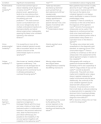
Fourth step: assessment of articular elbow pathologyIn the fourth step of the algorithm the clinician must examine if there is any articular pathology that could account for the symptoms experienced by the patient with intractable lateral epicondylitis. At this stage, the clinician will have already confirmed or ruled out a tendinopathy of the extensor muscles, the radio-humeral plica and the radial nerve as the causes of the patient's problems. The symptoms of snapping, blockage and/or apprehension can coexist with pain typically felt when loading the extensor muscles of the wrist, which commonly appears in epicondylalgia3,7. If the cause of intractable lateral epicondylitis is exclusively a tendon and/or neural pathology, these symptoms are not present and therefore if any of them are experienced, the clinician must suspect the presence of an articular dysfunction in the elbow13,33. In these cases, it is advisable to exclude the pathologies in Table 1 through the patient's physical examination and the use of imaging tests.
Differential diagnosis of intractable lateral epicondylitis if the patient has snapping, blockage and/or apprehension.
| Condition | Significant characteristics | Test19–20 | Considerations about imaging tests |
|---|---|---|---|
| Posterolateral rotary instability36–38 | It is the most common type of elbow instability and its diagnosis is mainly clinical36–38. In the physical exam, apprehension tends to be more obvious than subluxation or dislocation due to the patient's pain and protection37.The most common cause is a traumatism but it can also occur iatrogenically due to multiple corticosteroid injections to treat epicondylitis and lateral elbow surgery when inadequately repairing the lateral ulnar collateral ligament or the extensor tendons37. | Table-top relocation test.Stand-up test/chair push-up test.Push-up test.Lateral pivot shift test (awake/under anaesthesia)/posterolateral rotatory apprehension test)This is a highly specific test but it has a low degree of sensitivity with the patient awake7–38.Posterolateral drawer test. | Many patients have normal or subtly abnormal X-rays37.Although MRIs can be used, it must be borne in mind that injury to the lateral ulnar collateral ligament is not always easily identifiable in cases of chronic posterolateral rotary instability36.There is currently no consensus on the role of this test in the diagnosis of this ligament injury36.Dynamic fluoroscopy and ultrasounds can be useful if the diagnosis is confusing since they show ulnar head subluxation or ulnohumeral widening when either a posterolateral rotary drawer or a supination force on the elbow is applied34. |
| Varus posteromedial instability36 | It is caused by an injury to the lateral collateral ligament usually after a traumatism which can also produce a fracture of the anteromedial aspect or coronoid apophysis36. | Gravity-assisted varus stress test36. | The elbow varus stress test under anaesthetics is the diagnostic gold standard, an opening occurs in the ulnohumeral region36.There is currently no consensus on the role of ultrasounds in the assessment of the lateral ligament complex in relation to this instability36. |
| Valgus instability36,39 | Also known as “medial collateral ligament insufficiency”.This ligament can be damaged due to traumatism or repetitive elbow overuse and it is commonly found in athletes participating in overhead throwing sports39. | Moving valgus stress test.Valgus stress test/ligamentous instability test.Milking manoeuvre. | Arthrography with a saline or gadolinium-enhanced MRI has a sensitivity of 97% and a specificity of 100%36.Valgus stress X-rays can be useful39, as well as dynamic ultrasounds. They both can show medial joint instability when valgus stress is applied, assessing the medial ulnohumeral articular space. Additionally, the continuity of the ulnar collateral ligament can be observed with ultrasounds40. However, it must be borne in mind that in both tests an articular widening is observed which is expected in the dominant limb of overhead athletes, making outcome interpretation and diagnosis more difficult36. |
| Annular ligament injury33–35 | Some of these patients do not experience snapping or blockage.Patients with this injury can have a history of ulnar head fractures/luxation or malformations, fractures in the proximal radioulnar area, distal humeral fractures or prior elbow arthroscopy.It is possible to find instability in the annular ligament together with instability in the radioulnar joint35. | Symptoms may be caused by the gliding of the annular ligament on the ulnohumeral joint when bending and stretching the elbow35. | MRI or dynamic ultrasounds can help confirm the diagnosis35. |
Injuries to the radial collateral ligament and the lateral ulnar collateral ligament are frequent in patients with chronic epycondilalgia8,14. Significant elbow instability can be found with the use of the conventional physical exams detailed in Table 1. However, there may exist a type of subtle varus instability caused by an injury to the lateral elbow collateral ligament complex, which can be hard to detect in a conventional physical examination due to muscular restrictions. Research on elbow microinstability is scarce but there is evidence to support the inclusion of this clinical syndrome in the differential diagnosis of intractable lateral epicondylitis14. The clinician can rule out this subtle varus instability if no injury to the lateral ligaments is found in the imaging tests (with a negative predictive value of 98.7%). In contrast, if an abnormality is found in these structures, there is the possibility that the patient may have this microinstability. In a study by Kwak et al., 15 out of the 28 abnormal elbow MRIs examined presented subtle instability (with a positive predictive value of 53.6%). In consequence, in order to confirm this articular dysfunction, visualization of the radiocapitellar joint with the use of fluoroscopy is recommended in order to find a widening larger than 1.5mm at the joint when stressed under anaesthetic14. Additionally, it is important to underscore that these cases of frank or subclinical instability tend to compensate at muscular level causing Myofascial Pain Syndrome and Myofascial Trigger Points. These symptoms of muscular origin overlap with pain caused by the articular dysfunction, making the diagnosis even harder, and therefore muscular dysfunction must always be assessed when the clinician suspects any alteration in articular stability, as described in step five of the algorithm.
Moreover, in this fourth step the clinician must also exclude the presence of any injury to the annular ligament33,35 and of any intra-articular loose bodies in the elbow33,34, as shown in Table 1.
Fifth step: confirming or excluding Myofascial Pain Syndrome and Myofascial Trigger PointsIt is known that muscular dysfunction is involved in the chronic symptoms of patients with epicondylalgia16. In step five of the algorithm, the clinician will either exclude or confirm the presence of myofascial pain. The prevalence of Myofascial Pain Syndrome in lateral elbow pain is variable with a level of implication of the different muscles that varies according to different studies41–44. Although subjective and manual physical examination can be confusing, it is recommended that the clinician includes the following muscles in the differential diagnosis (Table 2).
Muscles whose Myofascial Trigger Points can be involved in intractable lateral epicondylitis and that the clinician must assess (from highest to lowest prevalence).
| - Extensor carpi radialis brevis muscle- Finger extensor muscle- Extensor carpi radialis longus muscle- Brachioradial muscle- Brachial triceps (mainly lateral fibres of the medial head)- Supinator muscle- Anconeus muscle- Extensor carpi ulnaris muscle |
Pain reproduction through the precise and analytical palpation of each muscle and/or the reproduction of symptoms with the use of dry needling can help the clinician to confirm this dysfunction as an element responsible for the symptoms16. The examination of each muscle under ultrasound control greatly improves the reliability of the assessment.
Sixth step: excluding the cervical spineIn the sixth step of the algorithm referred pain from the cervical spine must be excluded as the cause of chronic elbow pain7. In order to do this the clinician must answer the following questions: is it the neck or the elbow the region that reproduces the symptoms? Is there radicular pain? During the assessment, the clinician will ask the patient about the presence of concomitant neck pain and will assess the presence of any restriction in active range of motion of the neck and any neurological symptoms in the upper limb. During the patient's physical exam, the reproduction of lateral elbow pain through manual palpation and/or active and/or passive movements of the cervical or thoracic spine44–46 must alert the clinician as they can indicate the involvement of the spine in the patient's intractable lateral epicondylitis47,48.
Seventh step: examining central sensitization and psychosocial factorsCentral sensitization is involved in the pathophysiology of extensor tendon epicondylitis just as in other tendinopathies of the upper limb and in many musculoskeletal pain syndromes7,49 so its assessment will be the focus of the seventh and last step in this differential diagnosis algorithm. Anxiety and depression can also be frequently present in these patients17,50. Although further research is needed, the clinician is advised to consider psychosocial factors and include psychological assessment tools when dealing with patients with intractable lateral epicondylitis if necessary, once the rest of diagnoses have been confirmed or excluded.
DiscussionEpicondylalgia is the most common cause of lateral elbow pain in adults and when treatments do not seem to work, it is labelled “intractable lateral epicondylitis”. Does this clinical entity really exist or is it a misdiagnosis that consequently results in wrong treatment? The amount of structures that can cause the symptoms is really big and several clinical syndromes can coexist in lateral elbow pain. Moreover, the traditional tests used to diagnose epicondylalgia do not help in the screening of the different pathologies because many of them are positive and they are probably more useful as functional assessment parameters than as differential diagnostic tools. On the other hand, imaging tests alone are not enough for the diagnosis since most of the structures responsible for the symptoms in intractable lateral epicondylitis can be asymptomatic in spite of the fact that test images can show anatomo-pathological changes or the other way round: they can have a normal appearance and be symptomatic. That is why the clinician must be very meticulous when checking if there is any correlation between tissue changes and the patient's subjective and physical findings, which explains why a comprehensive and precise physical exam of the patient is essential and decisive for a correct clinical interpretation.
On the other hand, it must be taken into account that emotional and psychological factors can play a role in the perpetuation of symptoms. Nevertheless, there is the possibility that these are the consequence and not “the cause” of the pain and disability as it is normally claimed. For this reason the clinician must avoid attributing responsibility for pain chronification in patients with epicondylalgia to central sensitization and to psychosocial factors before making a comprehensive differential diagnosis of the different clinical syndromes that can coexist and get confused thus avoiding erroneous interpretations in the management of these patients51.
ConclusionIntractable lateral epicondylitis is a complex entity comprising different clinical syndromes that can coexist and get mixed up. That is why it is necessary to perform a systematic and comprehensive assessment of the patient like the one proposed in this article, where the musculotendinous articular and neural dysfunction responsible for the patient's symptoms is rigorously analyzed from highest to lowest prevalence. This seven-step differential diagnostic algorithm can be very useful for those clinicians dealing with these patients and can be base for future research, focusing on the identification of different subgroups of patients with intractable lateral epicondylitis for a correct clinical interpretation.
Conflict of interestThe authors declare that there is no conflict of interest.

