

Plantaris tendon is implicated in some cases of load-resistant Achilles tendinopathy. The tendon courses close to the medial Achilles tendon mid-portion prior to insertion onto the medial calcaneus, although there is variation in course and insertion. Clinical suspicion of plantaris involvement includes persistent medial Achilles pain unresponsive to a rehabilitation programme and imaging revealing a thickened plantaris tendon and/or focal medial Achilles tendinosis. Potential mechanisms include compression or shearing forces between the plantaris and Achilles tendons. Initial treatment should consist of a modified loading programme avoiding end-range loading. Resistant cases may be amenable to surgical excision of the plantaris demonstrating good clinical outcomes in the short and long term, although the evidence is limited to case series. Percutaneous methods show promise but require further evaluation.
El tendón del plantar delgado está implicado en algunos casos de tendinopatía de Aquiles resistente a la carga. El tendón se desplaza cerca de la porción media del tendón de Aquiles antes de su inserción en la región medial del calcáneo, aunque existen variaciones anatómicas en el curso y la inserción. La influencia del plantar delgado en la tendinopatía de Aquiles debe sospecharse ante un dolor en la región medial del Aquiles persistente y que no responde a un programa de rehabilitación y en el que las pruebas de imagen revelan un tendón plantar engrosado y / o tendinosos medial focal del tendón de Aquiles. Los mecanismos potenciales incluyen fuerzas de compresión o cizallamiento entre el tendón de Aquiles y el plantar delgado. El tratamiento inicial debe consistir en un programa de carga modificado que evite la carga en el tramo final. Los casos resistentes pueden ser susceptibles a la extirpación quirúrgica del plantar delgado que ha demostrado buenos resultados clínicos a corto y largo plazo, aunque la evidencia se limita a series de casos. Los métodos percutáneos son prometedores, pero se requieren más estudios que demuestren su validez real.
Chronic painful mid-portion Achilles tendinopathy is a common condition in running and jumping athletes but also in older non-athletic individuals.1 It represents the most common pathology among patients reporting persistent pain in the Achilles tendon region.2 Furthermore, it is one of the most common tendinopathy presenting in a general population cohort.3 As the aetiology and pathophysiology is not fully understood, treatment of this condition can be challenging.4 While most clinical cases improve with loading programmes, some patients are load-resistant and may be subjected to interventions with less efficacy and evidence-base. Recently, several authors have suggested that interference by the plantaris tendon may be implicated in some of these load-resistant cases.5–7
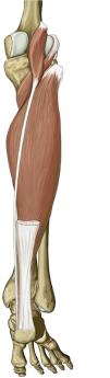
What is the plantaris tendon?The plantaris tendon arises from the plantaris muscle deep and medial to the lateral head of gastrocnemius.8 The tendon commences in the proximal calf and passes medial between the soleus and gastrocnemius9 and eventually courses close to the medial Achilles tendon mid-portion before commonly inserting onto the medial calcaneus (Fig. 1).9 Plantaris tendon course and insertion but also length and width can be highly variable between individuals.10 Cadaver studies on large specimen numbers found that most plantaris tendons insert onto the medial or anterior-medial aspect of calcaneus, but some insert directly onto the Achilles tendon or deep fascia.11,12 However, it can potentially terminate into all structures along its course.13

The variation in course and insertion may play an important role in the aetiology of mid-portion Achilles tendinopathy (see below). Although used as an important muscle in grasping objects with feet in primates, the plantaris is thought to be less functional in humans.9 The muscle is most active in plantarflexion when the knee is fully extended, but it contributes little to relative lower limb power compared to gastrocnemius or soleus muscles.14 However, due to its comparably large number of muscle spindles, it is thought to serve as proprioceptive organ for other plantar flexors.15
What is the evidence for implication in mid-portion Achilles tendinopathy?There is increasing clinical, imaging and morphological evidence implicating the plantaris tendon in some cases of persistent Achilles tendinopathy.
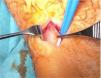
Several authors have implicated the plantaris tendon in clinical cases of intractable Achilles tendon pain. Steenstra and van Dijk5 were the first to report this clinical association in three cases proporting that some cases of persistent Achilles tendinopathy had Achilles and plantaris tendons that were closely associated and surrounded by inflamed tissue. Similarly, in a larger case series, Alfredson6 found that some cases of intractable Achilles tendinopathy were found to have a plantaris tendon closely adherent to the medial Achilles (see Fig. 2). In a few cases, the plantaris tendon was invaginated into the medial wall of the Achilles with richly vascularised fatty infiltration between the two tendons.6,16

Imaging has demonstrated a possible association between plantaris tendon and painful mid-portion Achilles tendinopathy. A recent study comparing macroscopic findings with clinical and imaging findings found that a high proportion of cases with suspected plantaris tendon involvement revealed a focal area of hypoechogenicity and increased blood flow in the medial Achilles.16 These focal changes in the medial Achilles corresponded to the position of the plantaris tendon during surgical excision. Using a novel imaging modality called Ultrasound Tissue Characterisation (UTC), focal areas of red and black echopixels representing disorganised matrix were found in the medial Achilles tendon in almost all cases. Interestingly all of these cases had tenderness in the medial aspect of the Achilles tendon.16
In addition, morphological studies have demonstrated an association between plantaris pathology and possible peripheral nociception. In a case series examining excised plantaris tendons from patients with clinically verified plantaris associated mid-portion Achilles tendinopathy, all specimens of excised plantaris tendons exhibited tendinosis-like morphological changes including disorganisation of collagen fibres, abnormal tenocyte morphology and increased tendon vascularity.17 In addition, immunohistochemical studies demonstrated sensory and sympathetic innervation within the plantaris tendon and in the peritendinous connective tissue between the Achilles and plantaris tendons supporting the hypothesis that these structures could play a role in nociception.18
What is the potential mechanism of plantaris-associated tendinopathy?While the mechanism of plantaris-associated Achilles tendinopathy remains unknown, recent observational and biomechanical studies have shed light on possible aetiological factors.
In normal individuals, the Achilles and plantaris tendons are positioned in the same paratenon and glide freely. Abnormal widths and/or thickening of the plantaris tendon in combination with close apposition of this tendon to the medial Achilles could lead to compressive19 and/or shearing forces20 leading to peritendinous inflammation or localised tendinopathy. Some authors suggest the inflammatory process may eventually result in a pertendinous adhesion onto the tendons restricting any further gliding.21 This hypothesis is supported by studies demonstrating multi-planar motion differential between the plantaris and Achilles tendons during passive plantarflexion and dorsiflexion22 and increased compressive forces between the two tendons at different ankle joint positions.23 In addition, the plantaris tendon has a higher intrinsic stiffness than the Achilles tendon in normal cadaveric specimens24 further supporting the potential for tethering of the plantaris against the Achilles tendon under tension.
As well as the differences in biomechanical properties, anatomical variations in the course and insertion of the plantaris tendon could contribute to the development of pathology. A recent study demonstrated a plantaris tendon in all 107 cadaveric specimens but they described difficulty in defining the plantaris tendon in some cases because the tendon was firmly adherent to the Achilles tendon.10 Furthermore, there was high variability in the course and insertion of the plantaris tendon with the tendon inserting onto 9 different locations. Moreover, certain anatomic variations could predispose to greater interference between the plantaris and Achilles tendon leading to greater compressive forces between the two tendons.21
In summary, biomechanical and anatomical factors of the plantaris tendon could lead to greater compression and/or shearing forces between the plantaris and Achilles tendons predisposing to Achilles tendinopathy. Further research is required to determine the importance of these factors in the development of plantaris-associated mid-portion Achilles tendinopathy.
What are the important diagnostic features of plantaris tendon involvement in mid-portion Achilles tendinopathy?There is a paucity of information regarding the clinical presentation or diagnostic features of plantaris-associated Achilles tendinopathy. Nonetheless, a few studies have attempted to define specific features based on clinical and imaging findings.
Clinical presentation of plantaris-associated tendinopathy has been described as load-related Achilles tendon pain situated primarily at the medial aspect of the Achilles tendon. In a recent study,16 which used surgical exploration to verify the close relation between the plantaris and Achilles tendons, most cases reported medial but rarely lateral Achilles pain and tenderness. Other authors have confirmed medial Achilles tendon pain as suggestive of plantaris tendon involvement.5,6 One case report described flicking or snapping in the medial Achilles.25Medial Achilles pain that is unresponsive to a comprehensive rehab programme may be suspicious for plantaris involvement and requires further evaluation and investigation.26
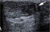
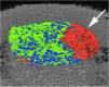
Variation in the course and insertion can render visualisation of the plantaris tendon difficult. Nonetheless, imaging can assist in the identification of the plantaris tendon and associated local pathology. A study using conventional 2D ultrasound and blood flow revealed a thickened plantaris tendon, focal medial Achilles tendinosis and increased blood flow in most cases of surgically-verified plantaris involvement see Fig. 3.16 In addition, using a novel imaging called Ultrasound Tissue Characterisation (UTC), all these cases demonstrated disorganised collagen structure represented by red and black echopixels in the medial Achilles tendon mid-portion (Fig 4), which is distinct from changes in usual mid-portion Achilles tendinopathy where they traditionally occur in the ventral aspect.16
How do you manage plantaris-associated mid-portion Achilles tendinopathy?

Most cases of mid-portion Achilles tendinopathy respond to a comprehensive and structured loading programme. Both eccentric and heavy slow resistance programmes26,27 have demonstrated effectiveness in reducing pain and improving function and return to sport in mid-portion Achilles tendinopathy. However, it is unknown whether plantaris-associated tendinopathy as a sub-group responds less well to loading programmes. Anecdotally, loading into dorsiflexion was thought to increase compression forces between the plantaris and Achilles tendons and reduce the responsiveness to loading programmes compared to cases without plantaris involvement. However, a recently published cadaveric study found that pressures were greatest at plantarflexion rather than dorsiflexion and lowest at a neutral ankle position challenging this assumption.23 A more effective programme for plantaris-associated mid-portion Achilles tendinopathy might involve heavy tendon loading into mid-range ankle positions, although further studies are needed to confirm clinical application of recent biomechanical studies.
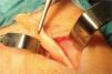
Surgical excision of the plantaris has been performed by various groups with good outcomes (see Fig. 5). Alfredson excised the plantaris tendon in patients with suspected plantaris involvement in mid-portion Achilles tendinopathy. Excision was performed under local anaesthetic and combined with a ventral scraping of the medial Achilles tendon.6 Most cases were satisfied with the treatment 12 months after the procedure,28 and a recent (but unpublished) study confirmed good outcomes in a long term follow up (2–13 years). Good outcomes have also been described by other groups using similar open methods7,29,30 and endoscopic approaches.31

The exact mechanism by which surgical excision has a positive clinical effect is unknown. Histopathological studies have demonstrated sympathetic sensory nerves within plantaris tendons and peritendinous fat between the plantaris and Achilles tendons.18 Therefore, excision of the plantaris tendon and scraping of the fat may remove sensory nerves and reduce nociceptive drive and pain perception. Alternatively, removal of the plantaris tendon might reduce shearing or compression forces on the medial Achilles and subsequent compression-induced tendinosis. This theory is supported by studies demonstrating improvement in tendon structure on UTC after plantaris tendon excision.29,32 Nonetheless, as effect of surgery may be related to other factors including a placebo effect, further higher level studies such as randomised controlled trials using a sham control are required to support the use of surgery in load-resistant plantaris cases.
Although these procedures are minimally invasive, plantaris tendon surgery exposes patients to certain risks, albeit small as 2% of cases developed infection or wound breakdown.2 Recently, a percutaneous US guided method has been proposed. Smith et al.33 confirmed successful incision of a plantaris tendon in twenty cadaveric specimens using US-guided percutaneous technique and local anaesthetic. Whether incision is enough to improve clinical outcomes or needs combination techniques including scraping or complete plantaris excision requires further analysis.
ConclusionPlantaris tendon is implicated in a subgroup of patients with mid-portion Achilles tendinopathy. Clinical and morphological studies point to the plantaris tendon as a possible patho-aetiological factor in some cases. A possible mechanism of interference is through compressive or shearing forces leading to peritendinous inflammation and/or compression-induced Achilles tendinopathy. Diagnosis is supported by findings of load-related medial Achilles tendon pain, localised medial Achilles hyperalgesia and ultrasound demonstrating an enlarged plantaris tendon and focal medial Achilles tendinosis. UTC can also assist in confirmation of diagnosis. Initial treatment should be conservative and involve heavy loading, and a recent biomechanical study suggests loading in mid-range might be more effective. Surgical excision of plantaris tendon has demonstrated improvement in clinical and imaging findings after plantaris tendon excision, but higher level studies are required to prove effectiveness. Percutaneous incision of plantaris tendon is a possible alternative to open excision, but clinical effectiveness needs evaluation.
Conflict of interestAuthors declare that they don’t have any conflict of interests.